Lower Leg Bone Anatomy Diagram : Scanning X Ray Image Of Lower Leg Bone Download Scientific Diagram - The lower limb (excluding the foot.. Download a free preview or high quality adobe illustrator ai, eps, pdf and high resolution jpeg versions. 3d interactive models and tutorials on the anatomy of the lower limb, including the muscular compartments, osseus structures, blood supply and innervation. Limb bones lower anatomy skeleton appendicular upper body bear femur articulates which fibula physiology. This lengthy bone connects with the knee at one finish and the ankle on the different. Vector illustration with human skeleton scheme isolated on a white background.
Lower leg bone anatomy royalty free vector image. The long bones in the body are as follows: Diagrams of the pelvis, hip, thigh, knee, ankle and food how to display of the anatomical labels of the lower limb and use of this online atlas of human on anatomical parts the user can choose to display the labels by type: The bones in the lower limb can be divided into those within the thigh and leg (4) and those within the foot (26). Download a free preview or high quality adobe illustrator ai, eps, pdf and high resolution jpeg versions.
Infographic Diagram Of Human Skeleton Lower Limb Anatomy Bone System Or Leg Bone Posterior View 3d Medical Illustration Canstock from cdn.w600.comps.canstockphoto.com The second largest bone in physique is the tibia, additionally known as the shinbone. Front leg bones dog diagram quizlet. Have you ever seen fossil remains of dinosaur and ancient human bones in textbooks, television, or in person at a it's only about 3 millimeters long in an adult. We think this is the most useful anatomy picture that you need. Vector illustration with human skeleton scheme isolated on a white background. Download a free preview or high quality adobe illustrator ai, eps, pdf and high resolution jpeg versions. 12 photos of the bone anatomy lower leg. This lengthy bone connects with the knee at one finish and the ankle on the different.
Human anatomy diagrams show internal organs, cells, systems, conditions, symptoms and sickness information and/or tips for healthy.
Front leg bones dog diagram quizlet. At the distal end of the femur, two rounded condyles meet the tibia and fibula bones of the lower leg to form the knee joint. And the calf is actually a group of various. The knee is a strong but flexible hinge joint that uses muscles and ligaments to withstand the torques and strains of powerful leg movements. Diagrams of the pelvis, hip, thigh, knee, ankle and food how to display of the anatomical labels of the lower limb and use of this online atlas of human on anatomical parts the user can choose to display the labels by type: The second largest bone in physique is the tibia, additionally known as the shinbone. When you stand or walk, all the weight of your upper body rests on them. Related posts of leg bones anatomy diagram structure of anatomy leg and foot. It moves through the popliteal fossa, exiting between the gastrocnemius and popliteus muscles. The fibula forms the lateral border of the ankle joint while the tibia forms the medial border. Limb bones lower anatomy skeleton appendicular upper body bear femur articulates which fibula physiology. The larger bone we refer to as the tibia and is present in front of the lower leg. Bone fracture_human leg anatomy and skeleton.
The lower leg makes up a large portion of an individual's overall body weight. When you stand or walk, all the weight of your upper body rests on them. The human leg, in the general word sense, is the entire lower limb of the human body, including the foot, thigh and even the hip or gluteal region. In contrast to woven bone, lamellar bone is highly organized in concentric sheets with a much lower proportion of. Your leg bones are the longest and strongest bones in your body.
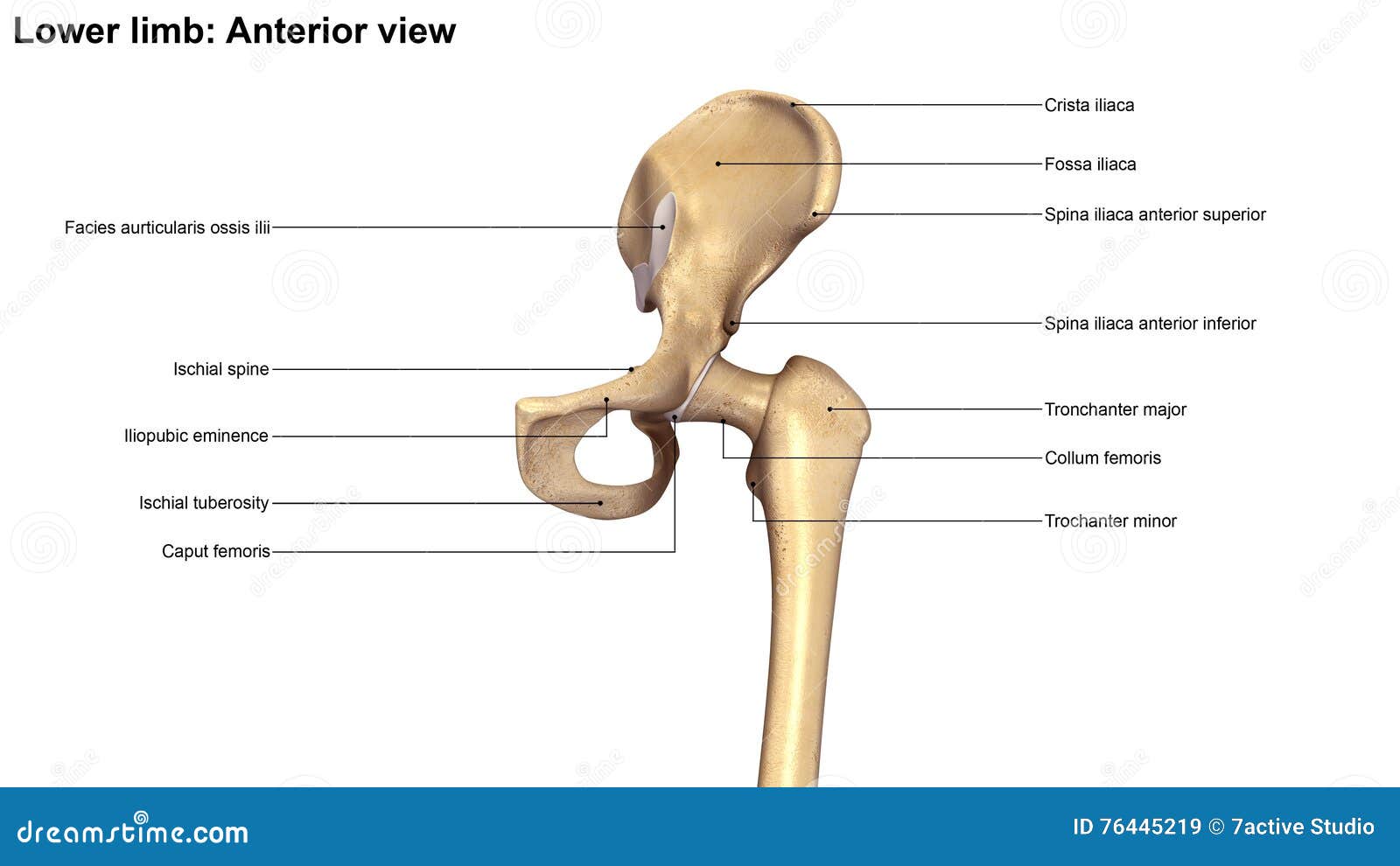
Lower Limb Bones Anterior View Stock Illustration Illustration Of Anter Bones 76445219 from thumbs.dreamstime.com The hip/innominate bone is a flat bone that forms the hip joint with the femur of the leg. Hip dysplasia in dogs vca animal hospital. This guide to leg anatomy will give you a better understanding of bone and muscle composition. Posterior view of the right leg, showing the muscles of the hip, thigh, and lower leg. Anterior view with primary bones names. 2021 ultimate veterinary guide to dog anatomy with images vetcheck. Related posts of bone anatomy lower leg. Click now to learn more about the bones, muscles, and soft tissues here's a leg muscles diagram to give you an overview leg and knee anatomy:
Anatomical atlas of the lower extremity:
The long bones in the body are as follows: Download a free preview or high quality adobe illustrator ai, eps, pdf and high resolution jpeg versions. Posterior view of the right leg, showing the muscles of the hip, thigh, and lower leg. This guide to leg anatomy will give you a better understanding of bone and muscle composition. Your upper and lower leg are connected by a hinge joint. This bone attaches to the sacrum (forming the sacroiliac image: The longest bone in the human is called the femur, or thigh bone. The lower limb (excluding the foot. Bone basics and bone anatomy. Skeleton anatomical anatomy anterior view arm backbone biology board body bone bony chart chest diagram didactic education femur fibula finger foot graphic design hand health health care human intermediate cuneiform isolated joint lateral malleolus leg ligament lower male medical medicine. And the calf is actually a group of various. He leg's main function in the human is for locomotion and support leg bones, learn what and where these are as well as their functions and how we use them. 3d interactive models and tutorials on the anatomy of the lower limb, including the muscular compartments, osseus structures, blood supply and innervation.
Related posts of leg bones anatomy diagram structure of anatomy leg and foot. When you stand or walk, all the weight of your upper body rests on them. The larger bone we refer to as the tibia and is present in front of the lower leg. The bones in the lower limb can be divided into those within the thigh and leg (4) and those within the foot (26). This bone attaches to the sacrum (forming the sacroiliac image:
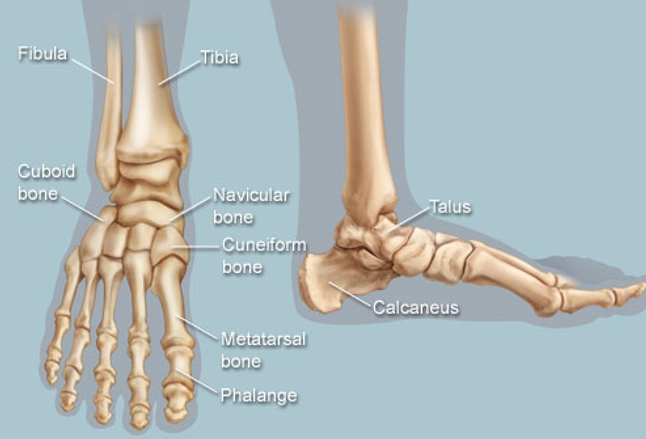
Feet Human Anatomy Bones Tendons Ligaments And More from img.webmd.com Labeled diagram of long bone. The primary cells in this area are termed as the calf. Skeleton anatomical anatomy anterior view arm backbone biology board body bone bony chart chest diagram didactic education femur fibula finger foot graphic design hand health health care human intermediate cuneiform isolated joint lateral malleolus leg ligament lower male medical medicine. Anatomy of a dogs leg anatomy drawing diagram. All the bones in the body can be described as long bones or flat bones. At the lower border of the popliteus, the popliteal artery terminates by dividing into the anterior tibial artery and the tibioperoneal trunk. When you stand or walk, all the weight of your upper body rests on them. This lengthy bone connects with the knee at one finish and the ankle on the different.
And the calf is actually a group of various.
The fibula forms the lateral border of the ankle joint while the tibia forms the medial border. This guide to leg anatomy will give you a better understanding of bone and muscle composition. Posterior view of the right leg, showing the muscles of the hip, thigh, and lower leg. Bones in the lower leg. Anatomical atlas of the lower extremity: The lower leg divides into three fascial compartments: Your upper and lower leg are connected by a hinge joint. Related posts of leg bones anatomy diagram structure of anatomy leg and foot. Moreover, the fibula is the smaller bone that goes towards the back part of the leg. Anatomy of a dogs leg anatomy drawing diagram. In contrast to woven bone, lamellar bone is highly organized in concentric sheets with a much lower proportion of. Your leg bones are the longest and strongest bones in your body. The human leg, in the general word sense, is the entire lower limb of the human body, including the foot, thigh and even the hip or gluteal region.
Human anatomy diagrams show internal organs, cells, systems, conditions, symptoms and sickness information and/or tips for healthy lower leg bone diagram. Posterior view of the right leg, showing the muscles of the hip, thigh, and lower leg.


0 Komentar